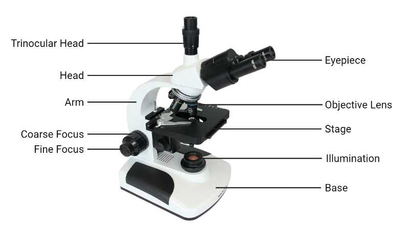
The exploration of intricate viewing instruments reveals a fascinating interplay of elements that enhance our understanding of the microscopic world. Each element plays a crucial role in magnifying and clarifying the objects under examination, providing insight into structures that are otherwise invisible to the naked eye.
By examining the various components, one can appreciate how these parts work in harmony to create a seamless experience for the user. From the initial collection of light to the final image projected for observation, each section contributes to the overall functionality, enabling detailed analysis and exploration of minute specimens.
Grasping the significance of these components not only aids in the effective use of the instrument but also enriches one’s knowledge of the science behind it. This understanding fosters a deeper appreciation for the technology that has advanced our ability to study the tiniest details of our universe.
Understanding Microscope Components
A thorough comprehension of the various elements that make up a viewing instrument is essential for effective usage and maintenance. Each section plays a crucial role in enhancing visibility and improving the overall functionality of the tool.
- Optical System: This area is responsible for the magnification and clarity of the image. It consists of different lenses that work together to provide a clear view of the subject being examined.
- Stage: This component serves as a platform where specimens are placed for observation. It often includes mechanisms for adjusting the position of the sample.
- Illumination Source: A light source illuminates the specimen, enabling details to be seen. This can be natural light or an artificial lamp.
- Base: The foundation supports the entire instrument, ensuring stability during use.
- Body Tube: This section connects the eyepiece and the optical system, allowing light to travel from the specimen to the viewer’s eye.
- Focus Mechanism: Essential for adjusting the clarity of the image, this feature allows users to refine their view of the subject.
Understanding these crucial components enhances the ability to utilize the instrument effectively, leading to better observations and more accurate results in various applications.
Essential Parts of a Microscope

Understanding the fundamental components of a viewing instrument is crucial for effective utilization. Each element plays a significant role in enhancing the clarity and magnification of the observed specimen, allowing for detailed examination. Familiarity with these key components can greatly improve the overall experience of scientific inquiry.
Key Components Overview
The following table highlights the primary elements, along with their functions, ensuring a comprehensive grasp of their importance in the functioning of the device.
| Component | Function |
|---|---|
| Objective Lens | Magnifies the specimen at various levels. |
| Eyepiece | Allows the viewer to see the magnified image. |
| Stage | Holds the specimen in place during observation. |
| Illuminator | Provides light to illuminate the specimen. |
| Focus Knob | Adjusts the clarity of the image. |
Conclusion
By comprehending the roles of these essential components, users can maximize the potential of the viewing instrument, leading to more productive scientific exploration. Mastery of these fundamentals is an invaluable asset in the pursuit of knowledge.
Functionality of Microscope Elements
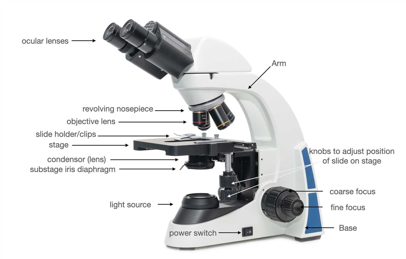
The intricate components of optical instruments play crucial roles in enhancing the visibility of minute details in various specimens. Each element contributes to the overall functionality, ensuring that users can observe and analyze with precision. Understanding these functions is essential for effective utilization and optimal results in scientific exploration.
Key Elements and Their Roles
Each component within the instrument serves a specific purpose, collaborating seamlessly to achieve clarity and focus. The various lenses amplify the image, while the lighting system illuminates the sample, revealing hidden structures that are otherwise imperceptible. Additionally, certain features enable adjustments to enhance the user’s experience and accuracy.
| Component | Function |
|---|---|
| Objective Lens | Magnifies the specimen and forms an initial image. |
| Condenser | Focuses light onto the specimen, improving illumination. |
| Eyepiece | Further magnifies the image for direct viewing. |
| Illuminator | Provides light source to enhance visibility of the sample. |
| Stage | Holds the specimen securely in place for observation. |
Collaboration of Elements
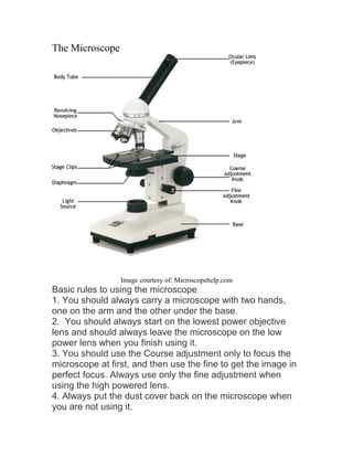
The harmonious interaction between these elements is fundamental for achieving high-quality observations. By adjusting various components, users can enhance clarity, contrast, and detail, facilitating a deeper understanding of the specimen. Mastery of these functionalities allows for proficient analysis in various scientific fields.
Detailed Labeling of Microscope Diagram
This section aims to provide a comprehensive examination of the components that make up a specific optical instrument used in scientific observation. By exploring the various elements and their functions, one can gain a deeper understanding of how this equipment operates and contributes to research and educational endeavors.
The first element is the support structure, which serves to stabilize the device while allowing for easy maneuverability. Next, the focusing mechanism plays a crucial role in enhancing the clarity of the observed specimen, enabling detailed examination. The light source illuminates the sample, ensuring that intricate features are visible. Additionally, the lenses are pivotal in magnifying the image, allowing for a closer inspection of minute details.
Moreover, the stage provides a platform for placing specimens, facilitating convenient access and observation. Each of these components interacts seamlessly, forming a cohesive unit that is essential for effective visual analysis. Understanding the function and relationship of these elements is fundamental for users aiming to utilize the instrument proficiently.
Types of Microscopes Explained
There are several types of optical instruments designed to magnify small objects, each offering unique capabilities and applications. Understanding these various instruments is crucial for selecting the right one for specific tasks, whether in a laboratory, classroom, or field study.
Common Categories of Optical Instruments
- Light Instruments: These utilize visible light and lenses to enlarge images of specimens.
- Electron Instruments: These employ electron beams instead of light, allowing for extremely high magnification and resolution.
- Digital Instruments: These integrate digital imaging technologies, making it easier to capture and analyze images.
Specialized Types
- Compound Variants: Typically used for thin samples, these utilize multiple lenses to enhance detail.
- Stereoscopic Models: Designed for three-dimensional viewing, these are ideal for examining larger objects.
- Fluorescence Variants: These instruments utilize fluorescence to visualize specific components within samples.
Importance of Each Microscope Part
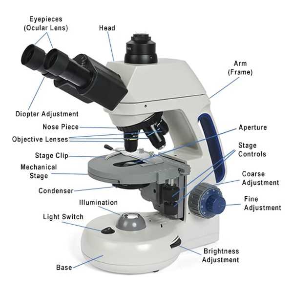
The various components of a viewing instrument play crucial roles in enhancing the overall functionality and effectiveness of the device. Each element contributes to the ability to observe intricate details of specimens, facilitating deeper understanding and exploration in the realm of science. Recognizing the significance of these elements aids users in maximizing their experience and outcomes.
Optical elements are essential for manipulating light to achieve clarity and precision in observations. These components determine how well details can be resolved, influencing the quality of images produced. A clear view enables users to study specimens in greater depth, fostering discoveries that may not be possible otherwise.
Mechanical features provide stability and support, allowing for precise adjustments during examinations. These mechanisms ensure that the focus and positioning of samples are accurate, which is vital for obtaining reliable data. Proper alignment of the instrument is necessary for effective observation, making these features indispensable.
Furthermore, illumination systems are critical in providing the necessary light for viewing specimens. The brightness and quality of light significantly impact the visibility of details, highlighting specific characteristics that are essential for analysis. Without adequate illumination, even the finest details may remain hidden, underscoring the importance of this element.
In summary, the unique contributions of each component enhance the instrument’s capability, making it a powerful tool for scientific inquiry. Understanding their roles allows users to appreciate the complexity and sophistication involved in the examination of microscopic structures.
How to Use Microscope Features
Utilizing the various components of a viewing instrument can enhance your observational experience significantly. Understanding how to manipulate these features effectively will enable you to achieve clearer images and better insights into your specimens.
Adjusting the Focus: Begin by using the coarse adjustment knob to bring your specimen closer to the lens. Once you have a basic view, fine-tune your focus with the fine adjustment knob to sharpen the image further. This method ensures that you can achieve optimal clarity for detailed examination.
Changing the Magnification: Most instruments come with multiple lenses of varying powers. To switch between magnifications, rotate the turret or nosepiece carefully until the desired lens clicks into place. This allows you to explore your specimen at different levels of detail, enhancing your overall understanding.
Illumination Settings: Proper lighting is crucial for effective observation. Adjust the diaphragm to control the amount of light entering the system. Increasing light intensity can improve visibility for transparent specimens, while reducing it may be beneficial for opaque samples.
Utilizing the Stage Controls: The stage clips are essential for securing your slides. Once in position, use the stage controls to move the slide around, allowing for a thorough examination of various areas of the specimen. This feature ensures that no detail goes unnoticed during your analysis.
Microscope Maintenance Tips
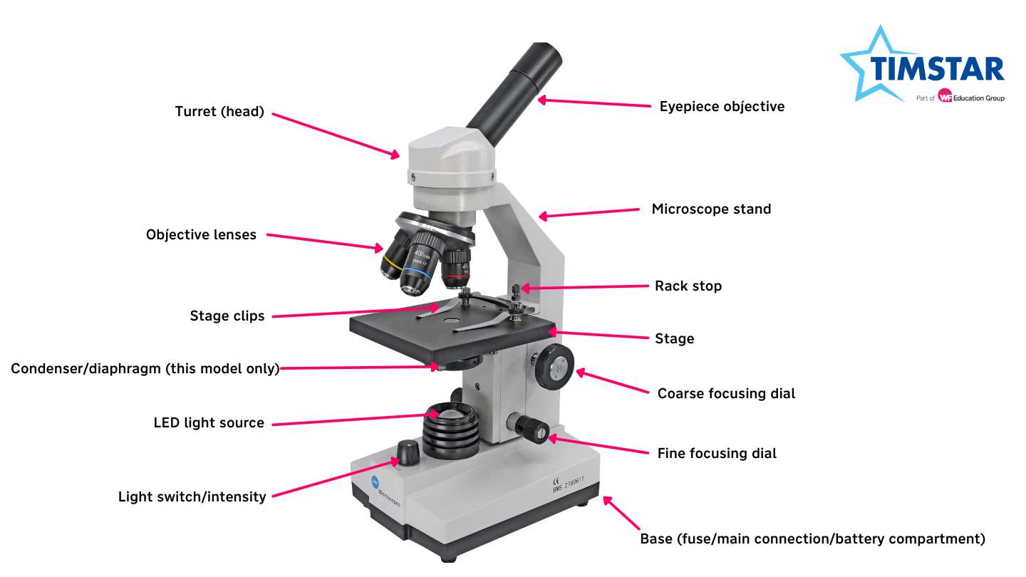
Proper care of optical instruments is essential to ensure their longevity and optimal performance. Regular maintenance helps prevent wear and tear, ensuring that users can achieve clear and precise observations over time. Implementing a few simple practices can significantly enhance the lifespan and functionality of these valuable tools.
First, always store the equipment in a clean, dry environment, preferably in a protective case when not in use. This minimizes exposure to dust and moisture, which can compromise the integrity of the lenses and other delicate components. Additionally, routinely check for any visible dust or fingerprints on the lenses, and clean them gently with appropriate materials to avoid scratches.
Regularly inspecting the mechanical parts for any signs of damage or misalignment is also crucial. If any issues are detected, seek professional assistance to address them promptly. Furthermore, ensure that all moving parts are adequately lubricated, which can help maintain smooth operation and prevent unnecessary wear.
Lastly, familiarize yourself with the manufacturer’s guidelines for upkeep and follow them diligently. By adopting these straightforward practices, you can maintain the quality and reliability of your optical instruments for years to come.
Common Issues with Microscope Parts
In the realm of optical instruments, various challenges can arise that may affect their performance. Understanding these common problems is essential for ensuring optimal functionality and extending the lifespan of these sophisticated tools. This section explores prevalent complications and their potential resolutions, facilitating a better user experience.
| Issue | Description | Solution |
|---|---|---|
| Focusing Problems | Difficulty in achieving a clear image can stem from misalignment or dirt on the lenses. | Regularly clean lenses and check alignment to ensure sharp focus. |
| Illumination Issues | Insufficient or uneven lighting can hinder visibility of the specimen. | Adjust the light source or replace bulbs as necessary to enhance illumination. |
| Stage Movement | Inability to smoothly move the stage may be due to wear or debris accumulation. | Inspect and clean the mechanical stage components, applying lubricant if required. |
| Dirty Components | Accumulation of dust and oil can obstruct clear viewing and affect overall performance. | Implement a regular cleaning schedule for all surfaces and internal mechanisms. |
Comparison of Microscope Models
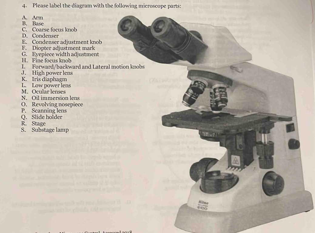
When exploring different optical instruments, it is essential to understand the various models available on the market. Each model presents unique features and capabilities that cater to specific applications and user needs. By examining the distinctions among these instruments, one can make informed choices based on functionality, design, and intended use.
Types of Instruments
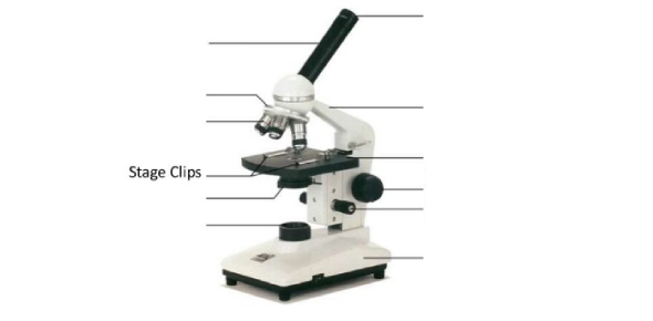
There are several types of optical devices, including compound, stereo, and digital variants. Compound instruments are commonly used in laboratories for detailed examination of thin specimens. Stereo models, on the other hand, offer a three-dimensional view, making them ideal for dissection and larger samples. Digital options incorporate technology to capture images and data, enhancing the research experience and enabling easier sharing of findings.
Features and Specifications
Different models vary significantly in terms of magnification, illumination, and stage size. Higher magnification ranges allow for more detailed observations, while advanced illumination systems, such as LED lights, improve visibility and reduce glare. Additionally, the size of the stage can affect the ease of sample placement and maneuverability, making it a crucial factor in selecting an appropriate model.
Applications of Microscopes in Science
The utilization of advanced optical instruments has revolutionized various scientific disciplines. These devices enable researchers to observe minute structures that are otherwise invisible to the naked eye, significantly enhancing our understanding of the microscopic world. The ability to magnify and illuminate objects at a cellular level has proven invaluable across multiple fields of study.
In biology, these tools play a crucial role in cellular research, allowing scientists to examine the intricacies of cells and their components. This examination aids in the discovery of cellular processes and interactions, which are fundamental to understanding life itself. Furthermore, advancements in imaging techniques have facilitated breakthroughs in genetics and microbiology, leading to innovations in medical research and treatment.
In materials science, these instruments are essential for analyzing the properties of materials at a granular level. Researchers can identify structural flaws, assess the composition of substances, and explore the characteristics of various materials, paving the way for the development of new technologies and applications. This level of analysis is crucial in industries ranging from nanotechnology to metallurgy.
Additionally, in environmental science, these tools enable the study of pollutants and microorganisms in various ecosystems. By observing the behavior and effects of these entities, scientists can develop strategies for conservation and pollution management. The insights gained from these observations contribute to our understanding of ecological balance and the impact of human activities on the environment.