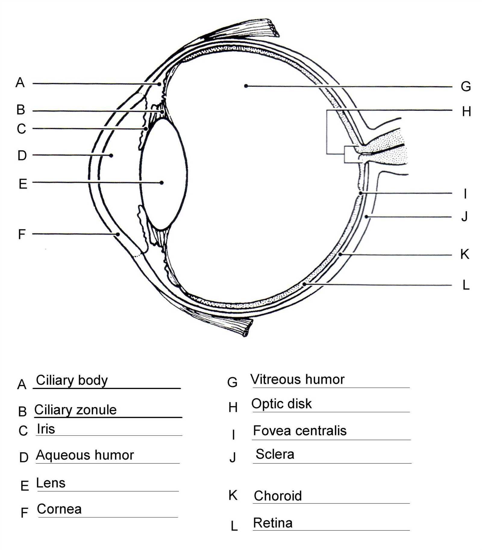
The complex system that facilitates vision is composed of numerous essential components, each contributing to the overall function of sight. By examining these elements, one can gain insight into how light is processed and converted into meaningful images. This exploration reveals the intricacies of human perception, allowing for a deeper appreciation of this remarkable sense.
Through a detailed illustration, individuals can familiarize themselves with various regions responsible for different aspects of vision. Each section plays a unique role, working harmoniously to enable clarity and focus. This educational approach provides a clear overview, assisting both students and enthusiasts in their quest for knowledge about ocular anatomy.
Moreover, recognizing these features enhances our understanding of common visual impairments and the importance of maintaining eye health. By becoming acquainted with these crucial elements, one can foster a greater awareness of the significance of proper care and attention. This foundation paves the way for informed discussions surrounding ocular science and advancements in vision correction.
Understanding Eye Anatomy
Delving into complex structure of visual organs reveals fascinating interconnections and functionalities. These intricate components work collaboratively to transform light into signals interpreted by brain, allowing us to perceive surroundings vividly. Gaining insight into this delicate framework enhances appreciation for remarkable processes enabling sight.
Key Components
Several essential elements contribute to overall functionality of visual organs. Each plays a crucial role, from capturing incoming light to processing images. Cornea serves as protective layer, while retina contains specialized cells that react to light. Additionally, lens adjusts focus, ensuring clarity of objects at various distances.
Functional Significance
Understanding significance of these intricate structures provides clarity on how visual information is processed. Pupil regulates amount of light entering, adapting to different lighting conditions. Optic nerve transmits signals from retina to brain, completing journey of visual processing. Each component, essential in its own right, contributes to holistic experience of sight.
Labeling the Eye Diagram
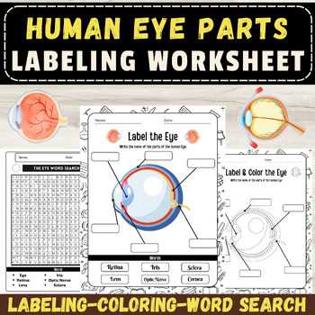
This section focuses on understanding various components of vision organs through a visual representation. Clear identification and proper naming of these elements are essential for grasping their respective functions and significance in the overall visual process.
By accurately marking each segment, learners can enhance their comprehension and retention of related concepts. Such exercises facilitate a deeper appreciation of how these structures work in harmony, contributing to the intricate mechanism of sight.
Utilizing labels not only aids in memorization but also encourages engagement with subject matter, making it easier to relate theoretical knowledge to practical applications. Emphasizing correct terminology fosters a foundation for further exploration into the fascinating field of ocular anatomy.
In summary, labeling serves as a vital tool for both educators and students, promoting an interactive learning experience while illuminating the complexities of visual perception.
Function of Eye Components
The intricate structure of vision relies on various elements that work together harmoniously to create a clear perception of the surroundings. Each component plays a vital role, contributing to the overall functionality necessary for sight. Understanding how these structures operate enhances our appreciation of the complexities involved in visual processing.
| Component | Function |
|---|---|
| Cornea | Provides initial focus and protection for the underlying structures. |
| Pupil | Regulates light entry by adjusting its size in response to brightness. |
| Iris | Controls pupil size and adds color to the visual organ. |
| Lens | Fine-tunes focus by altering shape, allowing for clear images at varying distances. |
| Retina | Transforms light into neural signals, initiating visual processing. |
| Optic Nerve | Transmits visual information from retina to brain for interpretation. |
Common Eye Disorders Explained
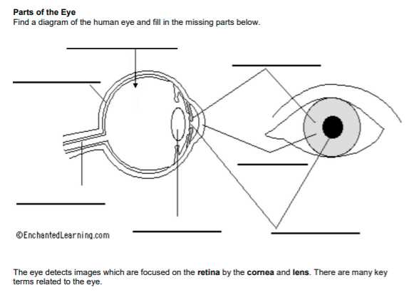
Visual health is essential for overall well-being, yet many individuals face various conditions that can affect their sight. Understanding these common ailments can empower individuals to seek timely intervention and adopt preventative measures. Awareness plays a crucial role in managing and treating such issues effectively.
Myopia, often known as nearsightedness, is a prevalent refractive disorder where distant objects appear blurry while close ones remain clear. This condition arises when light entering the optical system is focused incorrectly, typically due to an elongated shape of the eyeball.
Hyperopia, or farsightedness, is the opposite of myopia. Individuals with this condition struggle to see nearby objects clearly, while distant vision may be sharp. It occurs when the eyeball is shorter than normal, causing light rays to focus behind the retina.
Astigmatism results from an irregular curvature of the cornea or lens, leading to distorted or blurred vision at all distances. This imperfection can cause visual discomfort and difficulty focusing, often requiring corrective lenses.
Presbyopia is a natural age-related condition where the ability to focus on close objects diminishes. It typically becomes noticeable in the early to mid-40s as the lens loses elasticity, making reading or other close-up tasks challenging.
Cataracts develop when the lens becomes cloudy, resulting in blurred vision, glare, and difficulty seeing at night. This condition is often associated with aging but can also result from other factors such as diabetes, smoking, or prolonged exposure to sunlight.
Glaucoma encompasses a group of conditions that damage the optic nerve, often linked to increased pressure within the ocular structure. This progressive condition can lead to vision loss if left untreated, making regular screenings vital for early detection.
Being informed about these common visual disorders encourages individuals to prioritize regular check-ups and seek professional guidance when experiencing symptoms. Proactive management can significantly improve quality of life and preserve visual function.
The Role of the Cornea
The cornea serves as a vital structure within visual perception, acting as the initial barrier to light. This transparent layer is crucial for focusing light rays onto the retina, ensuring that images are sharp and clear. Its unique curvature and refractive properties significantly influence overall vision quality.
Light Refraction
One of the primary functions of this clear shield is light refraction. As incoming light passes through, it bends, altering its path toward the lens. This process is essential for achieving optimal focus, allowing the brain to interpret images accurately. Without proper refraction, clarity diminishes, leading to distorted perception.
Protection and Health
Beyond focusing capabilities, this transparent layer also plays a protective role. It safeguards underlying structures from environmental damage, including harmful particles and pathogens. In addition, it contributes to overall health by maintaining moisture and facilitating healing processes. When compromised, vision can be adversely affected, highlighting its importance in maintaining visual integrity.
Importance of the Retina
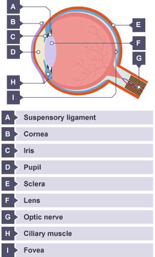
The retina plays a crucial role in visual perception, acting as a sensitive layer that captures light and transforms it into neural signals. This intricate process allows individuals to perceive their surroundings in vivid detail and color. Understanding the significance of this specialized tissue is essential for appreciating how vision functions and maintaining overall ocular health.
Functions of the Retina
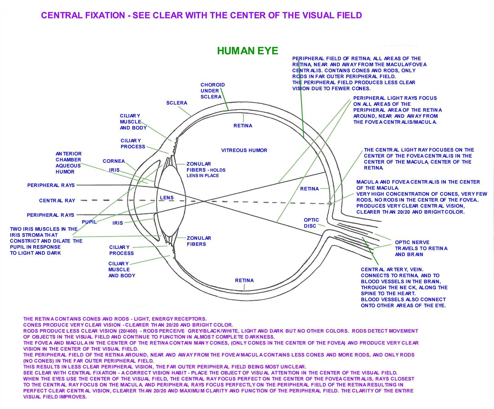
- Light Sensitivity: It detects and responds to various light intensities, enabling clear vision in different lighting conditions.
- Color Perception: Specialized cells within the retina facilitate the recognition of a wide spectrum of colors, enriching visual experience.
- Image Processing: It processes visual information before transmitting it to the brain, contributing to efficient recognition of objects and movement.
Common Disorders Affecting the Retina
- Retinal Detachment: A serious condition where the retina separates from its underlying support tissue, leading to potential vision loss.
- Diabetic Retinopathy: A complication of diabetes that affects blood vessels in the retina, causing vision impairment.
- Macular Degeneration: An age-related condition that deteriorates the central part of the retina, impacting detailed vision.
Preserving retinal health is vital for maintaining optimal visual function. Regular eye examinations and prompt attention to any visual changes can help safeguard this essential tissue.
How the Lens Works
This section explores functionality of a crucial component responsible for focusing light, enabling clear vision. It plays a significant role in adjusting focus depending on distance, allowing for sharp images to form on the retina.
Light rays entering this optical structure undergo refraction, changing direction as they pass through. This process is vital for bending incoming light, ensuring it converges correctly. Flexibility of this structure permits it to change shape, a mechanism essential for focusing on objects at various distances.
| Distance | Lens Shape | Focusing Ability |
|---|---|---|
| Far objects | Flatter | Less curvature |
| Near objects | Rounder | Increased curvature |
Muscles surrounding this optical element contract or relax, facilitating shape alteration. As these muscles tighten, the structure becomes thicker and more rounded, enhancing its power to focus on nearby items. Conversely, when these muscles relax, it flattens, optimizing focus for distant views.
Significance of the Iris
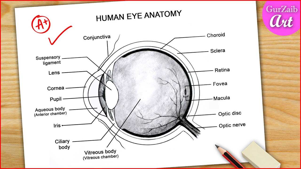
The iris serves as a crucial element within visual perception, playing a vital role in regulating light intake and enhancing clarity. This unique structure adapts dynamically to varying lighting conditions, ensuring optimal function and protection for surrounding components.
Functions of the Iris
- Light Regulation: Adjusts pupil size to control illumination entering.
- Color Variation: Offers distinct hues, contributing to individuality and aesthetic appeal.
- Health Indicator: Changes in coloration or pattern may signify underlying health conditions.
Impact on Vision
A well-functioning iris not only enhances visual acuity but also protects delicate structures from excessive brightness. Its ability to adapt swiftly to different environments enables clear sight in diverse conditions.
Vitreous Humor and Its Function
The vitreous humor plays a crucial role in maintaining structural integrity and supporting functions within the visual organ. This gel-like substance occupies the majority of the internal space, contributing to overall shape and providing a medium for light transmission.
Key functions of vitreous humor include:
- Providing cushioning and protection against mechanical shock.
- Maintaining intraocular pressure, essential for proper shape and function.
- Allowing light to pass through to the retina without obstruction.
- Serving as a reservoir for nutrients and waste products, aiding in metabolic processes.
Additionally, vitreous humor plays a significant role in supporting retinal health. It connects to the retina at various points, helping to stabilize its position while allowing for slight movements during eye motion. Changes in this gel can lead to various visual disturbances, highlighting its importance in visual function.
Exploring the Optic Nerve
Delving into intricate connections within visual processing reveals a crucial pathway that transmits signals from photoreceptors to various regions of the brain. This vital structure plays a pivotal role in interpreting what is perceived, contributing significantly to how organisms interact with their surroundings.
Structure and Function
This unique conduit consists of millions of nerve fibers, originating from retinal ganglion cells. These fibers converge to form a compact bundle, ensuring efficient transmission of visual information. Key functions include:
- Transmitting visual signals to the brain.
- Facilitating communication between different visual areas.
- Contributing to reflexive responses to visual stimuli.
Clinical Significance
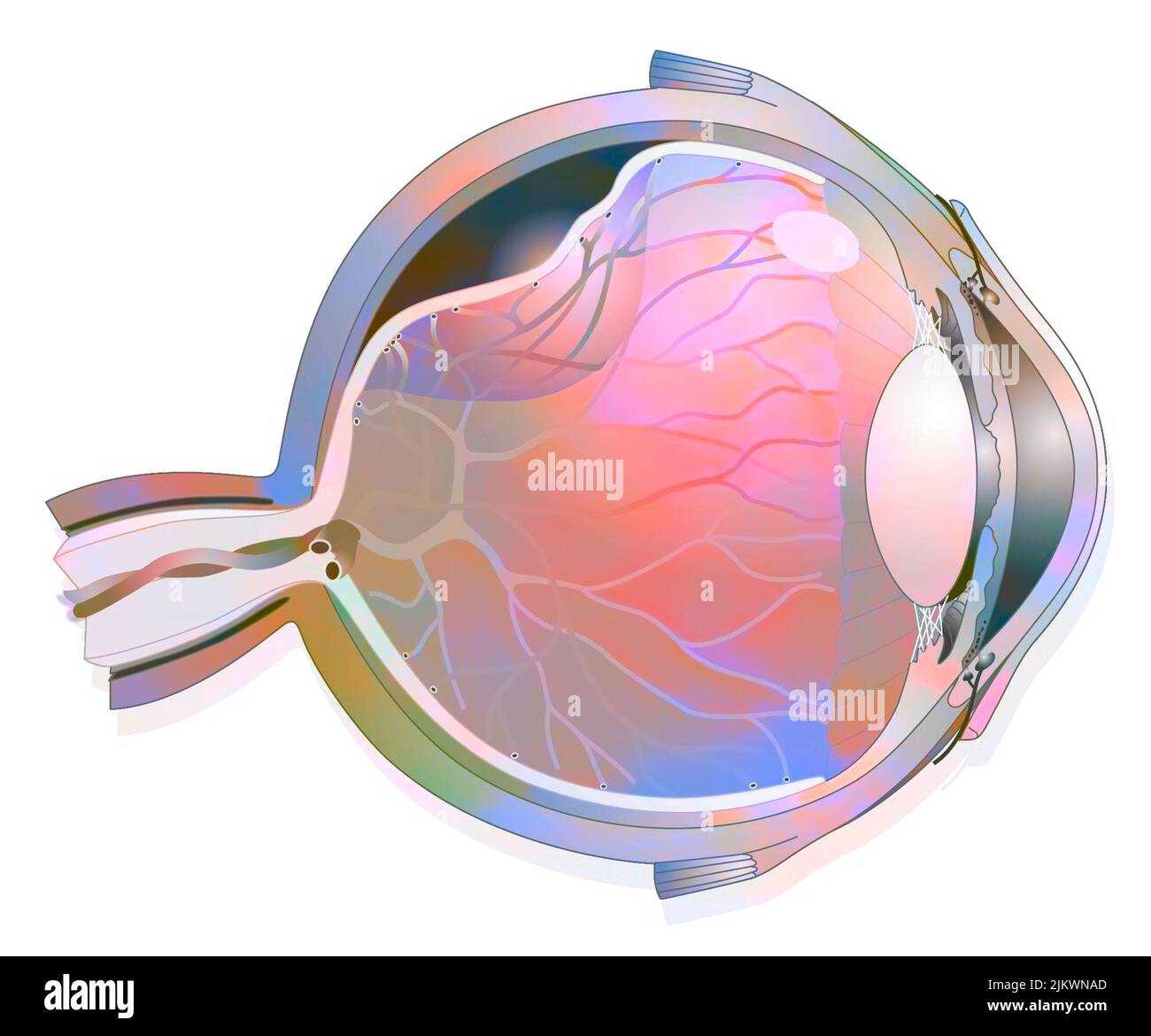
Understanding the anatomy and function of this conduit is essential for diagnosing and treating various visual disorders. Common issues include:
- Optic neuritis, which can lead to vision loss.
- Glaucoma, causing damage to the optic pathway.
- Compression from tumors, affecting signal transmission.
Investigation of this critical structure continues to enhance knowledge about visual perception and associated pathologies, underscoring its importance in both health and disease.
Protective Structures of the Eye
Maintaining clarity and function of visual organs is crucial for overall well-being. Various mechanisms and barriers work in harmony to shield these delicate structures from environmental threats, injuries, and harmful agents. Understanding these safeguards is essential for appreciating how vision is preserved.
External Barriers

The first line of defense comprises several external features that prevent dust, debris, and microorganisms from entering sensitive areas. Eyelids serve as movable covers, offering protection during rest and reacting reflexively to potential dangers. Additionally, lashes trap particles, while tear fluid maintains moisture and flushes away irritants.
Internal Defenses
Inside, multiple layers contribute to safeguarding against internal damage. Conjunctiva, a thin membrane, lines the inner surfaces, providing lubrication and further preventing foreign bodies from causing harm. Furthermore, specialized cells produce tears containing antimicrobial substances, enhancing the immune response and promoting healing.