
The human body is an intricate structure composed of numerous elements that work together to provide support, mobility, and protection. This framework serves as the foundation for our physical form, enabling a wide range of movements and functions essential for daily life. Each component plays a crucial role in maintaining the overall integrity and functionality of the organism.
To gain a deeper appreciation of this fascinating construct, it is important to explore the various elements that contribute to its design. From the large, weight-bearing structures to the delicate connectors, each plays a pivotal role in ensuring that the body operates efficiently. This exploration will illuminate how these elements interconnect, revealing the complexity and elegance of the human body.
By examining the composition and arrangement of these crucial features, we can better understand their significance and how they contribute to our overall health and mobility. A closer look at these integral components will provide insight into the mechanisms that support our physical capabilities and the ways in which they interact with one another.
Skeletal System Overview
The framework of the human body plays a crucial role in providing structure, support, and protection to vital organs. It serves as a foundation that enables movement while also facilitating various physiological processes. This intricate assembly consists of numerous interconnected elements, each contributing to the overall stability and functionality of the body.
Composed of a complex network of bones and connective tissues, this structure not only shapes the body’s form but also interacts with muscles to produce movement. The framework is also essential for producing blood cells and storing minerals, highlighting its multifunctional importance. Understanding this assembly is key to appreciating how it supports and sustains life.
Major Bones and Their Functions
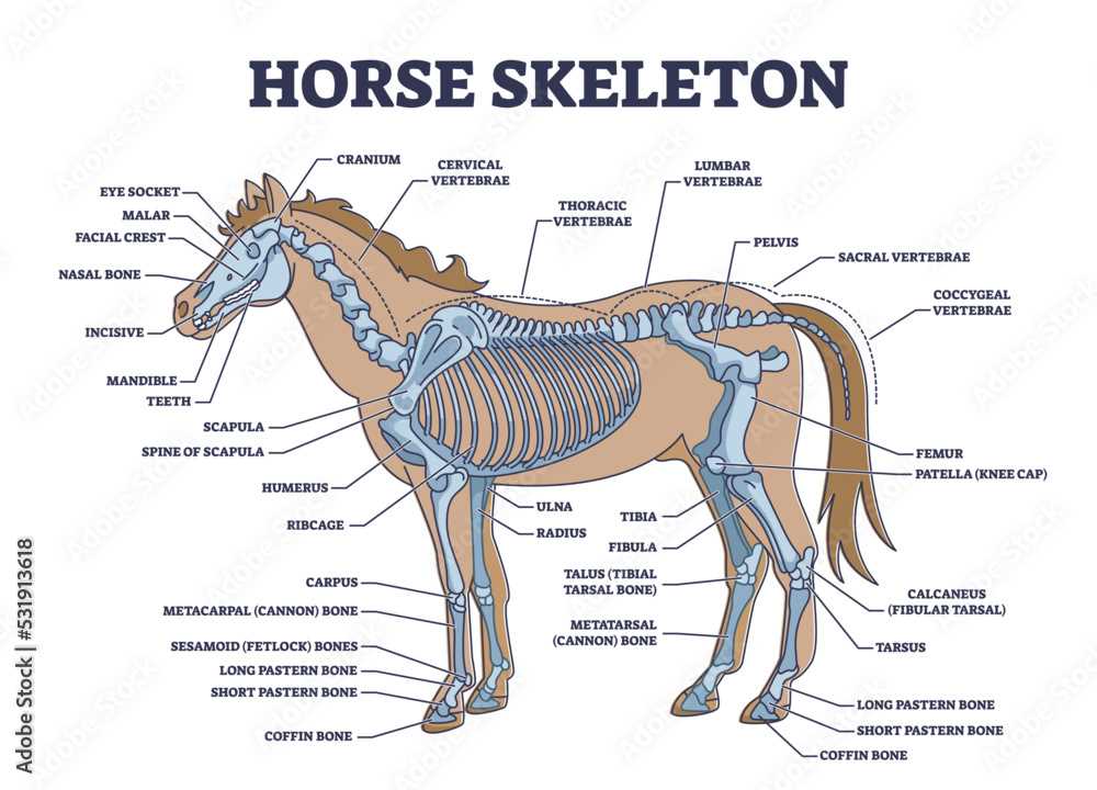
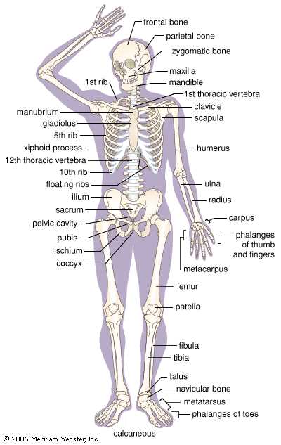
The human body is supported by a complex framework of hard structures, each serving crucial roles in maintaining overall health and functionality. Understanding these vital components is essential for appreciating how our bodies operate.
Here are some of the most significant bones and their respective functions:
- Skull
- Protects the brain from injury.
- Supports the structures of the face.
- Facilitates the process of eating and breathing.
- Spine
- Provides structural support and stability to the upper body.
- Protects the spinal cord, which is critical for nerve signal transmission.
- Enables flexibility and movement.
- Rib Cage
- Encases and safeguards vital organs like the heart and lungs.
- Facilitates breathing by expanding and contracting.
- Offers attachment points for muscles involved in respiration.
- Humerus
- Supports the upper arm and connects the shoulder to the elbow.
- Facilitates a wide range of arm movements.
- Femur
- The longest bone in the body, providing support for walking and running.
- Acts as a key component of the hip and knee joints.
- Tibia
- Contributes to the stability of the lower leg.
- Supports weight-bearing activities.
Each of these bones plays a distinct role in ensuring mobility, protection, and overall functionality of the body. Their interconnected nature highlights the intricate balance required for healthy physical activity.
Components of the Axial Skeleton
The axial framework serves as the central support structure for the body, playing a vital role in maintaining posture and protecting critical organs. This framework is composed of several key elements that work together to provide stability and strength. Understanding these components is essential for grasping the overall function of the body’s structural integrity.
Primarily, this framework consists of the vertebral column, which is composed of individual bones called vertebrae that are stacked to form a flexible yet sturdy column. This arrangement allows for both support and movement, enabling the body to bend and twist while safeguarding the spinal cord. The ribs, which encase the thoracic cavity, provide a protective barrier for vital organs such as the heart and lungs, while also facilitating respiratory movements.
Additionally, the skull, housing the brain, is an intricate structure composed of numerous bones that protect the central nervous system and support the facial framework. These various components not only contribute to the overall shape and form of the body but also play critical roles in numerous physiological functions.
In summary, the axial framework comprises several essential elements, each contributing uniquely to the body’s stability, protection, and functionality. An understanding of these components is crucial for appreciating how they collectively maintain the body’s integrity.
Understanding the Appendicular Skeleton
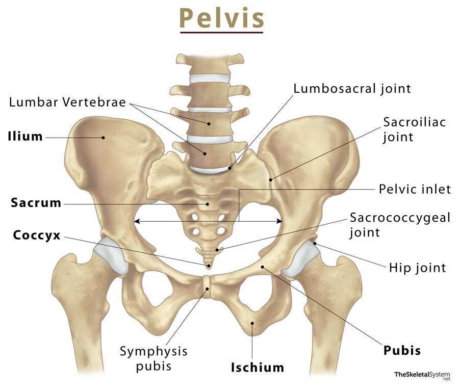
The appendicular region of the human framework plays a vital role in movement and functionality. It encompasses the structures that allow for a wide range of motions and activities, facilitating interactions with the environment. This section explores the key components and their significance in overall mobility.
Components of the Appendicular Framework
The appendicular region includes several key structures, which can be categorized into upper and lower divisions:
- Upper Division:
- Shoulder girdle
- Arms
- Wrists
- Hands
- Lower Division:
- Pelvic girdle
- Legs
- Ankles
- Feet
Functionality and Movement
The structures within this region enable various forms of movement, which are essential for daily activities. The following are key functionalities:
- Locomotion: Facilitates walking, running, and jumping.
- Manipulation: Allows for grasping, lifting, and holding objects.
- Balance and Posture: Contributes to stability and body alignment.
Understanding the components and functions of the appendicular region provides insight into its importance in daily life and physical activities.
Types of Joints in the Body
The human body boasts a remarkable variety of connections that facilitate movement and flexibility. These unique junctions play a crucial role in enabling a wide range of motions, contributing to the overall functionality of the organism. Understanding these different forms of articulation helps to appreciate how they support various activities, from simple tasks to complex athletic performances.
Joints can be categorized based on their structure and function. Synovial joints are the most common type, characterized by a fluid-filled cavity that allows for extensive movement. Examples include the knees, elbows, and shoulders. Cartilaginous joints, on the other hand, provide limited movement and are connected by cartilage, such as those found between the vertebrae in the spine.
Lastly, there are fibrous joints, which are connected by dense connective tissue and typically allow for very little movement. These are found in areas like the skull, where the bones are tightly bound to protect the brain. Each type of joint serves a distinct purpose, collectively contributing to the intricate design of the human anatomy.
Bone Tissue and Its Structure
Bone tissue serves as a fundamental component within the framework of the human body, providing both strength and support. This specialized connective tissue is crucial for various functions, including movement, protection of vital organs, and mineral storage. Understanding its composition and architecture is essential to grasping how these elements contribute to overall functionality.
At a microscopic level, bone tissue is composed of cells, fibers, and an extracellular matrix that collectively form its structure. The primary cell types include osteoblasts, responsible for bone formation; osteocytes, which maintain bone tissue; and osteoclasts, involved in resorption. These cells operate in harmony to ensure the tissue remains healthy and responsive to physiological demands.
The extracellular matrix is rich in collagen fibers, providing tensile strength, and a mineralized component primarily made of hydroxyapatite, which gives bones their hardness. This unique combination allows bone tissue to withstand compressive forces while remaining lightweight. The overall arrangement of these elements varies between different types of bone, such as compact and spongy bone, each serving distinct functions within the body’s architecture.
Furthermore, the organization of bone tissue is intricately linked to its functionality. The dense structure of compact bone forms the outer layer of bones, providing strength and support. In contrast, spongy bone, characterized by a network of trabecular structures, facilitates flexibility and reduces overall weight while maintaining structural integrity. This dual organization ensures that the framework of the body can endure various mechanical stresses encountered in daily activities.
Common Skeletal System Disorders
Various conditions can affect the framework of the body, leading to discomfort, pain, and mobility issues. Understanding these ailments is essential for effective prevention and treatment. Many of these disorders arise due to a combination of genetic factors, lifestyle choices, and environmental influences. This section will explore some of the most prevalent issues encountered in the human body’s structural framework.
Types of Common Disorders
Some conditions manifest through inflammation, degeneration, or malformation, significantly impacting an individual’s quality of life. It is crucial to recognize the symptoms and seek appropriate care to mitigate the effects of these disorders.
| Condition | Description | Symptoms |
|---|---|---|
| Osteoporosis | A condition characterized by decreased bone density, making bones fragile and more susceptible to fractures. | Back pain, loss of height, and fractures that occur easily. |
| Arthritis | An inflammatory condition affecting joints, causing pain and stiffness. | Swelling, tenderness, and reduced range of motion in affected areas. |
| Scoliosis | A curvature of the spine that can develop during growth spurts. | Uneven shoulders, back pain, and noticeable curvature of the spine. |
| Fractures | Breaks in bones resulting from trauma, stress, or underlying conditions. | Severe pain, swelling, and inability to use the affected limb. |
Conclusion
Awareness and early detection of these conditions can lead to better management and improved outcomes. Regular check-ups and a healthy lifestyle play crucial roles in maintaining overall health and well-being.
Role of Cartilage in Joints
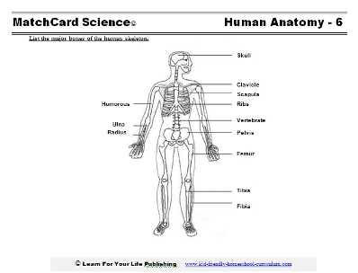
Cartilage serves as a crucial element in the functionality and stability of the connections between bones. Its unique properties allow it to withstand pressure while providing a smooth surface for movement. This specialized tissue plays a pivotal role in ensuring that the interactions between adjoining structures remain efficient and pain-free.
Key functions of cartilage include:
- Shock Absorption: It cushions the impact during physical activities, reducing the stress transferred to the underlying bone.
- Facilitating Movement: The smooth texture of cartilage allows for effortless gliding of bones against one another, promoting fluid motion.
- Providing Structure: Cartilage maintains the shape of certain structures, such as the ears and nose, while supporting the joints.
- Reducing Friction: The presence of this tissue minimizes friction between bone surfaces, preventing wear and tear over time.
Furthermore, the maintenance and health of cartilage are vital for overall joint functionality. Damage or degradation of this tissue can lead to discomfort and limited mobility, emphasizing the importance of protecting and caring for it through proper nutrition and lifestyle choices.
Growth and Development of Bones
The process of bone formation and maturation is essential for maintaining the structure and function of the human body. This intricate journey begins in early life and continues through adolescence, marked by significant changes influenced by genetics, nutrition, and physical activity. Understanding these phases can provide insights into how our bodies grow and adapt over time.
Phases of Bone Development
Bone development occurs in several distinct stages, each characterized by unique cellular activities and morphological changes. These stages include the initial formation of cartilage, its subsequent ossification, and the ongoing remodeling that occurs throughout life.
| Stage | Description | Key Processes |
|---|---|---|
| Embryonic | Initial formation of the cartilaginous model of bones. | Chondrogenesis, matrix production |
| Childhood | Progressive ossification where cartilage is replaced by bone tissue. | Ossification, mineralization |
| Adolescence | Growth plates gradually close as individual matures, ceasing height increase. | Epiphyseal closure, remodeling |
Influences on Bone Growth
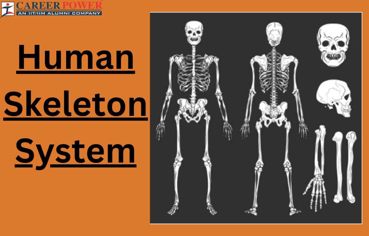
Various factors play a crucial role in bone growth and health. Nutritional intake, particularly calcium and vitamin D, significantly affects bone density and strength. Additionally, physical activity stimulates bone formation and remodeling, promoting overall skeletal health. Hormonal changes during puberty also accelerate growth, underscoring the complex interplay between biological processes in bone development.
Importance of Calcium in Bone Health
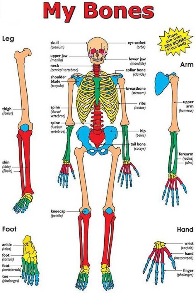
Calcium plays a crucial role in maintaining strong and healthy bones throughout an individual’s life. This essential mineral is not only vital for bone structure but also contributes to various physiological functions. Adequate calcium intake is necessary to prevent conditions related to bone density, ensuring longevity and overall well-being.
Role of Calcium in Bone Development
During the developmental stages of life, especially in childhood and adolescence, calcium is fundamental for the growth and maturation of bone tissue. Sufficient calcium levels during these critical periods lay the foundation for optimal bone density and strength in adulthood.
Consequences of Calcium Deficiency
A lack of calcium can lead to weakened bones, increasing the risk of fractures and conditions such as osteoporosis. It is essential to recognize the signs of calcium deficiency early to implement dietary changes or supplements to safeguard bone integrity.
| Calcium-Rich Foods | Calcium Content (mg) |
|---|---|
| Dairy Products (Milk, Yogurt) | 300 |
| Leafy Greens (Kale, Broccoli) | 150 |
| Fortified Foods (Cereals, Juices) | 200 |
| Fish with Bones (Sardines, Salmon) | 400 |
| Nuts and Seeds (Almonds, Chia Seeds) | 250 |
Diagnostic Techniques for Skeletal Issues
Identifying problems related to the body’s framework is crucial for effective treatment and recovery. A variety of methods are utilized to assess the condition and functionality of the bones and joints. These techniques aid in pinpointing the underlying causes of discomfort or mobility restrictions, facilitating informed decisions for medical intervention.
Among the most common approaches are imaging methods that provide visual insights into the structural integrity of the framework. Each technique offers unique advantages, enabling healthcare professionals to tailor assessments based on individual patient needs.
| Technique | Description | Uses |
|---|---|---|
| X-ray | A quick imaging technique that captures images of internal structures using radiation. | Detecting fractures, dislocations, and abnormalities in bone density. |
| Magnetic Resonance Imaging (MRI) | A non-invasive imaging method using magnetic fields and radio waves to create detailed images. | Evaluating soft tissues, cartilage, and joint conditions. |
| Computed Tomography (CT) Scan | A specialized imaging technique that combines X-ray images taken from different angles to create cross-sectional views. | Providing a comprehensive view of complex fractures and joint issues. |
| Bone Scintigraphy | A nuclear imaging technique that detects changes in bone metabolism. | Identifying infections, tumors, or unexplained pain. |
| Ultrasound | A non-invasive technique using sound waves to visualize soft tissues and fluid. | Assessing joint effusions and guiding injections or aspirations. |
Exercises to Strengthen the Skeleton
Engaging in physical activities can significantly enhance the integrity and durability of our internal framework. These exercises not only fortify the bones but also improve overall health, balance, and mobility. A well-rounded routine can lead to better posture, reduced risk of injuries, and a higher quality of life.
Weight-Bearing Activities
Incorporating weight-bearing exercises is crucial for maintaining bone density. These activities encourage the growth of robust tissues, making the structure less susceptible to fractures.
- Walking: A simple yet effective way to promote strength in the lower limbs.
- Running: This high-impact activity increases the load on bones, fostering growth.
- Dancing: Fun and engaging, dancing improves coordination and supports structural strength.
- Stair climbing: An excellent method for enhancing leg strength and endurance.
Resistance Training
Utilizing resistance can further stimulate the growth of dense tissues. By challenging the muscles, these exercises contribute to a resilient framework.
- Weightlifting: Targeted lifting of weights can build muscle and support the surrounding structures.
- Bodyweight exercises: Movements such as push-ups, squats, and lunges utilize one’s weight for resistance.
- Resistance bands: These versatile tools allow for varied intensity and can be adjusted for different fitness levels.