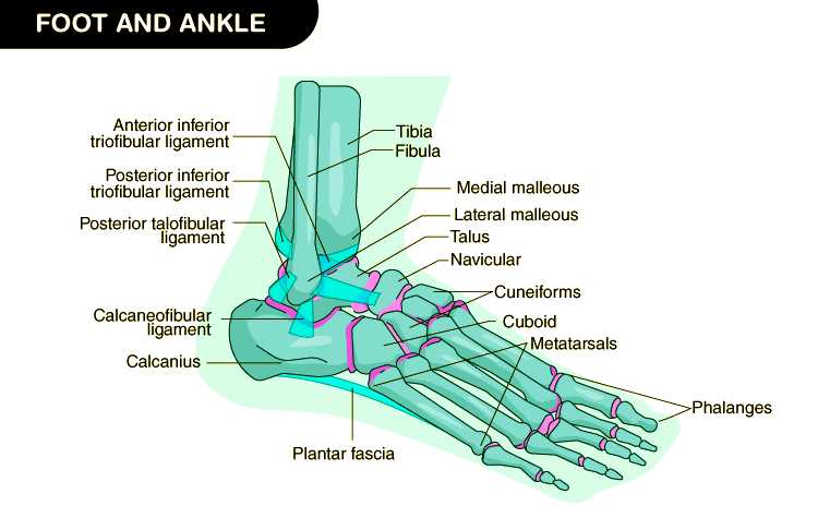
Exploring the complex arrangement beneath the ankle reveals an intricate network essential for movement and balance. Every component works in harmony, forming a stable foundation for walking, running, and standing. This section offers insights into the structural connections that ensure both mobility and support.
The lower extremity includes various segments, each contributing uniquely to posture, weight distribution, and shock absorption. These interconnected regions play vital roles in locomotion, adapting to uneven surfaces and dynamic forces with remarkable efficiency.
A closer look at these elements can deepen our appreciation for the biological design responsible for maintaining stability. Such knowledge proves valuable not only for medical professionals but also for anyone seeking to improve performance, prevent injuries, or understand everyday functionality better.
Anatomy Overview of Human Lower Limb Structure
The human lower extremity is a complex arrangement of bones, muscles, tendons, and ligaments that collectively support weight, enable locomotion, and maintain balance. This section provides an insightful exploration into the intricate architecture underlying human bipedalism.
Structure and Function
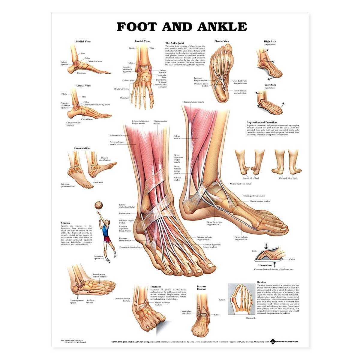
At its core, the lower limb comprises robust skeletal components seamlessly integrated with dynamic soft tissues. These elements work synergistically to facilitate mobility and transmit forces essential for daily activities. Key anatomical landmarks, including joints and muscular attachments, play pivotal roles in ensuring both stability and flexibility.
The arrangement of fibrous connective tissues, reinforced by specialized tendons and ligaments, serves to anchor muscles and govern joint movements. These structural adaptations contribute to the remarkable versatility observed in human ambulation, accommodating various terrains and physical demands.
Exploring further into this domain illuminates the profound adaptability and resilience embodied within the human foot and its associated anatomical framework.
Main Bones in the Foot Structure
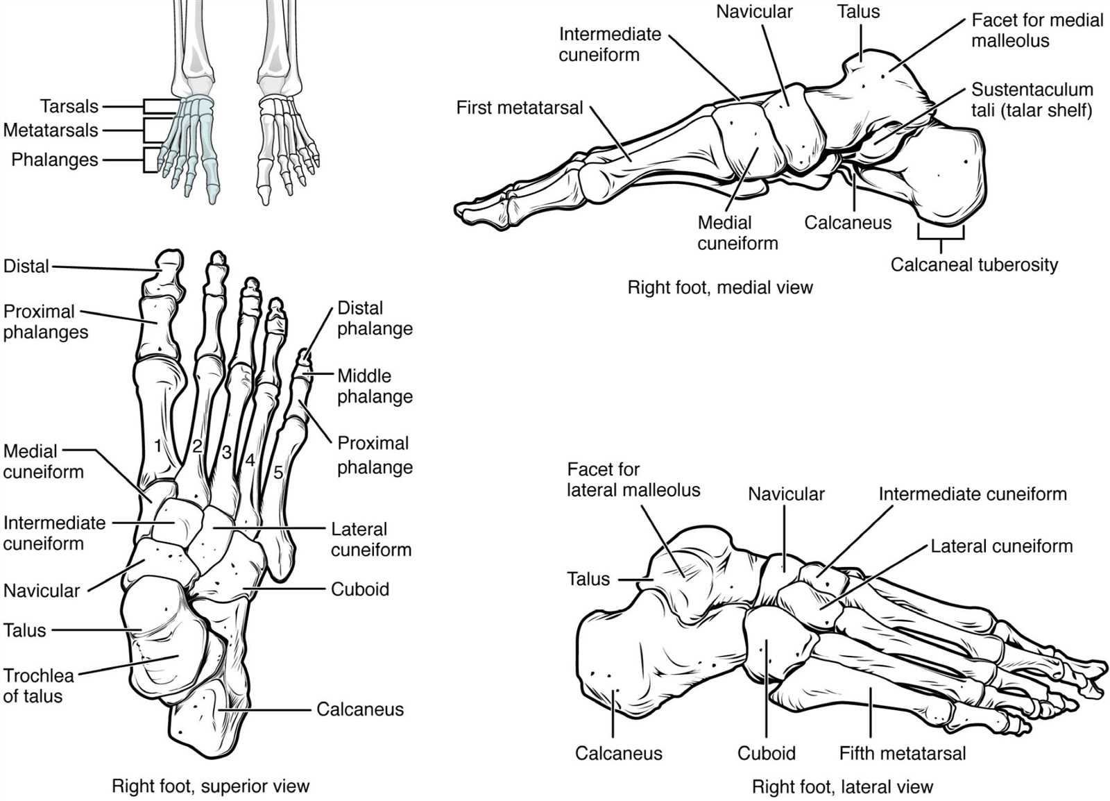
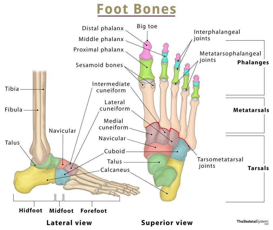
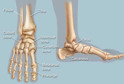
The skeletal framework at the base of the leg provides both stability and mobility, ensuring proper balance and weight distribution during movement. This intricate design supports everyday activities such as walking, running, and standing.
Several key bones work in harmony to shape this structure. These include larger elements responsible for absorbing impact and smaller components that enhance flexibility. Together, they form a unified system that adapts to various surfaces and conditions.
Each section plays a distinct role, from maintaining arch integrity to facilitating smooth transitions between steps. A solid understanding of these elements is essential for recognizing how injuries or conditions may affect locomotion.
The harmony between rigid components and flexible joints ensures the necessary balance between strength and agility, allowing seamless motion across different terrains.
Muscles Supporting Foot Movement
This section highlights how various muscle groups contribute to the intricate mechanics required for walking, running, and balancing. These tissues work together to create stability, mobility, and agility, making efficient movement possible.
- Intrinsic Muscles: Small muscles within the sole area that help maintain posture and provide fine control.
- Extrinsic Muscles: Larger groups originating from the leg, controlling more extensive movements like plantarflexion and dorsiflexion.
- Flexor Group: Enables curling actions and maintains grip strength on uneven surfaces.
- Extensor Group: Facilitates lifting movements, assisting in toe extension and preventing tripping.
- Abductor and Adductor Muscles: Responsible for spreading and drawing the toes together, aiding in balance adjustments.
The collaborative function of these muscle sets ensures proper coordination during each phase of motion, reducing strain and enhancing overall performance.
Ligaments and Tendons in the Foot
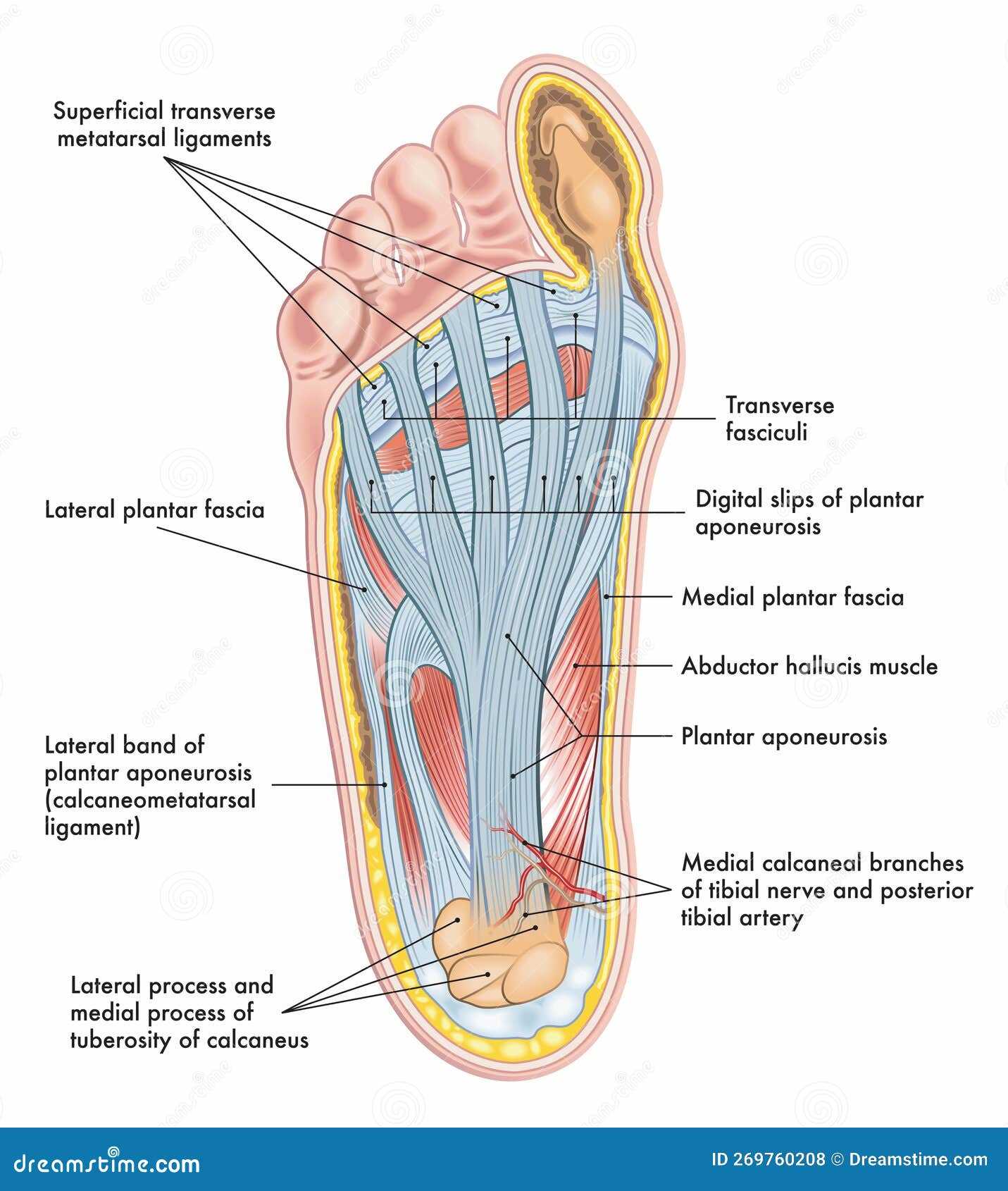
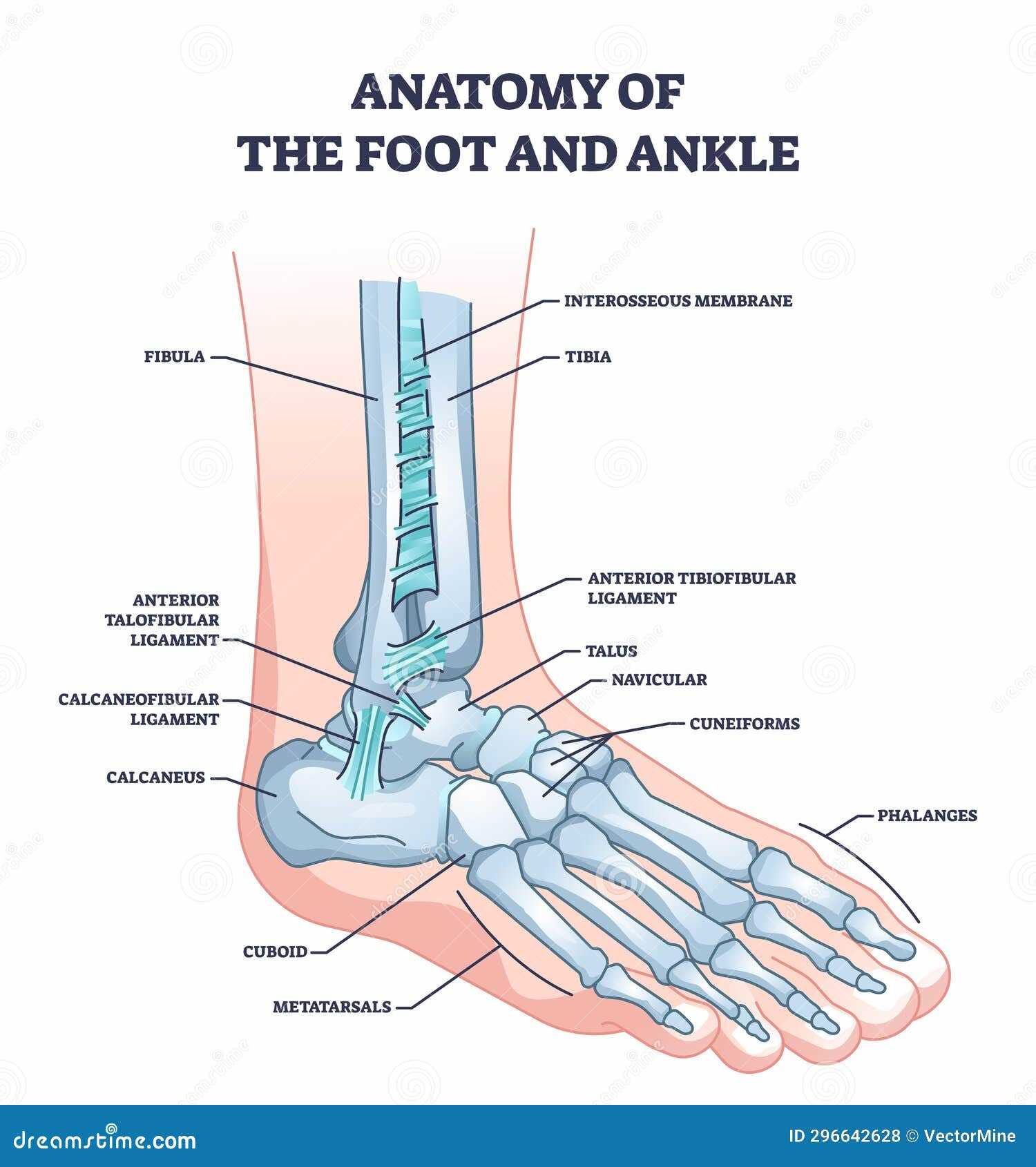
Within the lower extremity, flexible bands and connective tissues play essential roles in ensuring stability, mobility, and shock absorption. These structures interlink bones, muscles, and joints, contributing to balance and effective locomotion.
- Ligaments: These fibrous bands connect bones and are crucial for joint stability. They prevent excessive movement, reducing the risk of injury.
- Tendons: Tendons attach muscles to bones, transmitting forces necessary for movement. Their elasticity helps absorb impact during dynamic activities.
- Plantar Region: Contains key ligaments and tendons supporting the arch and providing cushioning during walking.
- Ankle and Heel Area: Strong ligaments and tendons ensure proper alignment, aiding in pivoting and weight distribution.
- Toes: Smaller yet vital tendons enable precise movements required for balance and grip.
Maintaining these tissues’ health is crucial for mobility, as they must withstand significant strain and tension during daily activities.
Blood Circulation and Foot Arteries
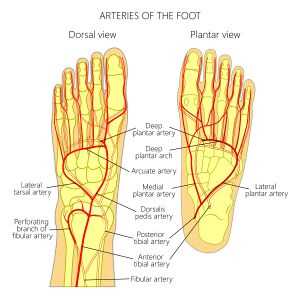
Efficient blood flow ensures proper nourishment and oxygen delivery throughout the lower extremities. Circulatory pathways in this region play a key role in maintaining mobility, stability, and overall health. Any disruption in these channels can affect vital processes, leading to discomfort or serious conditions.
Main Arterial Network
The primary arteries travel down the leg, branching into smaller vessels that reach the lower regions. These divisions supply crucial areas with fresh, oxygen-rich blood, supporting muscle function and skin integrity. Proper circulation helps maintain tissue health and allows for quick recovery from minor injuries.
Importance of Healthy Circulation
A well-functioning vascular system prevents the buildup of waste products and promotes healing. Issues such as poor circulation can result in fatigue, swelling, or more severe complications if not addressed promptly. Regular physical activity and maintaining balanced pressure on the lower limbs are essential for optimal vascular health.
Nerve Pathways Through the Foot
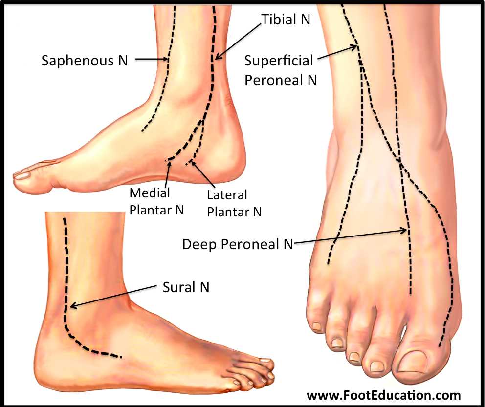
Understanding how signals travel through complex structures can offer valuable insights into motion control and sensory feedback. This section explores neural routes that govern movement, detect pressure, and manage coordination, ensuring smooth interaction between different areas.
Main Branches and Routes
- The primary neural channels control voluntary movement, enabling precise actions such as flexion and extension.
- Additional fibers relay tactile sensations, helping detect textures and subtle changes in surface contact.
- Certain branches also manage pain responses and temperature perception, protecting the body from harm.
Connections Supporting Mobility
- A network of smaller pathways communicates with joints, regulating balance and alignment.
- These channels also synchronize with muscles, ensuring a coordinated response to shifts in weight and position.
- Neural integration with higher centers allows rapid adjustments during motion, improving stability and posture.
Each neural pathway plays a role in maintaining fluid movement and sensory awareness, contributing to overall agility and well-being.
Arch Types and Their Function
Understanding various arch forms is essential for appreciating their roles in supporting body weight and enabling movement. Each type contributes uniquely to overall stability and mobility, playing a vital role in physical activities and daily tasks.
Types of Arches
- Normal Arch: Characterized by a balanced curve, this form provides adequate support and shock absorption.
- High Arch: This type has a pronounced curve, often leading to increased pressure on the ball and heel areas.
- Flat Arch: With minimal curvature, this form can result in overpronation, impacting alignment and function.
Functions of Different Arch Forms
- Weight Distribution: Each arch type helps distribute body weight evenly, reducing strain on specific areas.
- Shock Absorption: Curvature aids in absorbing impact forces during walking, running, and jumping.
- Stability: Arches enhance balance, allowing for efficient movement and prevention of injuries.
Heel Components and Their Role
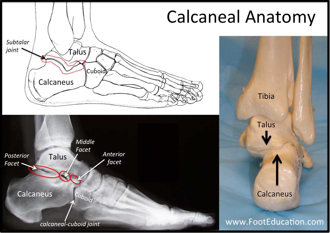
Understanding the anatomy located at the back of the lower limb is essential for appreciating its function and significance in overall mobility. This section will explore key structures contributing to stability, weight distribution, and movement efficiency in this specific area.
Structural Elements
Key structures in this region include bones, ligaments, and soft tissues. These components work together to provide both support and flexibility. The robust framework formed by the bones ensures durability, while the ligaments facilitate connections, allowing for controlled movements and maintaining stability.
Functionality
These anatomical features play a vital role in absorbing shock during activities such as walking, running, or jumping. They also contribute to balance and posture, helping to prevent injuries. Enhancing the understanding of these components can aid in addressing various conditions that may arise, leading to improved well-being and performance.
Toes Structure and Flexibility
Understanding the anatomy and adaptability of digits at the end of limbs is essential for appreciating their role in balance and mobility. These small yet significant components contribute to overall stability while allowing a range of movements crucial for various activities.
Each digit consists of bones, joints, and surrounding soft tissues, providing both strength and agility. The bones, known as phalanges, are organized into three sections in most digits, enhancing flexibility and enabling diverse movements. This arrangement allows for a natural curling and stretching motion, essential for actions such as gripping or pushing off surfaces.
Flexibility is further supported by ligaments and tendons, which connect muscles to bones, facilitating smooth movement. The intricate network of these structures ensures that digits can adapt to different surfaces, aiding in shock absorption during activities like walking or running. The ability to flex and extend freely is vital for maintaining a balanced posture and efficient locomotion.
Moreover, the skin covering these digits is designed to provide protection while allowing for sensitivity. This combination of structural elements and adaptability makes digits indispensable for performing daily tasks and engaging in physical activities.
Common Foot Joints and Connections
This section explores essential articulations and linkages within the lower extremity, highlighting their significance in mobility and stability. Understanding these structures is crucial for recognizing how they contribute to overall functionality during various activities.
Several key joint types exist, each playing a specific role in movement and load-bearing. Below is a summary of some of the major connections found in this region.
| Joint Name | Location | Function |
|---|---|---|
| Talocrural Joint | Between tibia, fibula, and talus | Facilitates dorsiflexion and plantarflexion |
| Subtalar Joint | Between talus and calcaneus | Enables inversion and eversion |
| Metatarsophalangeal Joints | Connecting metatarsals to phalanges | Allows flexion, extension, and some rotation |
| Interphalangeal Joints | Between adjacent phalanges | Permits flexion and extension |
Skin Layers and Protection Mechanisms
The outer covering of human anatomy serves crucial functions, including safeguarding against environmental threats and maintaining overall health. This protective layer comprises several strata, each with specific roles in defense and sensory perception.
Understanding these layers is essential for recognizing how they contribute to resilience and adaptability in various conditions. Below are the main components and their functions:
- Epidermis: The uppermost layer, primarily responsible for barrier functions and containing melanocytes for pigmentation.
- Dermis: Situated beneath the epidermis, this layer provides structural support, housing blood vessels, nerves, and various glands.
- Hypodermis: The deepest layer, acting as insulation and shock absorption while connecting skin to underlying structures.
Protection mechanisms are integral to maintaining integrity and function. Key features include:
- Barrier Function: The outer layer prevents pathogens and harmful substances from penetrating.
- Regeneration: Rapid healing processes enable recovery from minor injuries and abrasions.
- Sensory Reception: Specialized nerve endings in these layers detect temperature, pressure, and pain, contributing to reflexes and overall awareness.
In conclusion, the various layers of skin and their associated mechanisms play a pivotal role in defense, health, and sensory feedback, underscoring their importance in human anatomy.
Foot’s Role in Balance and Posture
Stability and alignment play a crucial role in overall movement and health. An intricate system of structures contributes to maintaining equilibrium, allowing individuals to perform various activities with confidence and ease. Understanding how these components interact can lead to better practices for enhancing physical stability.
Proper alignment is essential for distributing weight evenly across the body. This distribution helps prevent discomfort and injuries, promoting overall well-being. Additionally, strength and flexibility in specific areas can further improve stability during different activities.
The following table outlines key factors contributing to balance and posture:
| Factor | Description |
|---|---|
| Muscle Strength | Robustness in lower limb muscles aids in supporting weight and maintaining upright positions. |
| Joint Flexibility | Range of motion in joints allows for adaptive movements, reducing the risk of falls. |
| Nerve Sensitivity | Enhanced sensory feedback helps the brain coordinate movements and adjust posture effectively. |
| Surface Interaction | Different surfaces can affect stability; adapting to these changes is vital for maintaining balance. |
Maintaining optimal function in these areas can lead to improved posture and a reduced likelihood of injuries, thereby enhancing overall quality of life.